

1. Introduction
Knee osteoarthritis is a common degenerative musculoskeletal condition characterised by chronic pain, functional limitation, inflammation, and progressive loss of joint cartilage. With an ageing population and increasing sedentary lifestyles, this condition has become one of the leading causes of physical disability in adults and older individuals.
Given the clinical complexity of osteoarthritis, integrative therapeutic approaches involving non-invasive technologies and functional rehabilitation strategies have gained significant attention. Among these approaches, the combination of therapeutic laser (photobiomodulation), microcurrent therapy (MENS), and kinesiotherapy has shown promising clinical outcomes in reducing pain, modulating inflammation, supporting tissue regeneration, and improving functional mobility.
The purpose of this article is to present the scientific foundations, clinical evidence, and practical protocols for the combined application of these three modalities in managing knee osteoarthritis. All recommendations are based on the technologies currently in use at Bruno Physical Rehabilitation.
2. Scientific Basis of Combined Therapies
2.1 Therapeutic Laser (Photobiomodulation Therapy – PBMT)
Therapeutic laser therapy, also known as photobiomodulation therapy (PBMT), is a clinically validated, non-invasive treatment that uses specific wavelengths of light (typically within the red to near-infrared spectrum, ranging from 600 to 1,100 nm) to stimulate tissue repair, modulate inflammation, and relieve pain. This technology harnesses the biological effects of light at the cellular level, promoting homeostasis and regeneration.
The primary mechanism involves the absorption of light photons by chromophores within the mitochondria, particularly cytochrome c oxidase. This interaction accelerates the electron transport chain, leading to increased production of adenosine triphosphate (ATP), the cell’s energy currency. Enhanced ATP levels improve cell metabolism and viability, which is critical in tissues affected by osteoarthritis (OA), where hypoxia and metabolic dysfunction are common.
In addition, PBMT has been shown to:
- Promote angiogenesis and improve local microcirculation
- Modulate the expression of pro-inflammatory cytokines (e.g., IL-1β, TNF-α)
- Enhance fibroblast proliferation and collagen synthesis
- Stimulate chondrocyte activity and reduce cartilage degradation
These effects are dose-dependent and influenced by parameters such as wavelength, power output (mW), energy density (J/cm²), and treatment time. Research has suggested that an optimal energy density for knee OA ranges between 4 and 12 J/cm² per session [1].
A double-blind placebo-controlled study by Alayat et al. (2017) demonstrated significant improvement in pain scores and physical function in patients treated with PBMT, compared to placebo and exercise-only groups [2].
2.2 Microcurrent Therapy (Microcurrent Electrical Neuromuscular Stimulation – MENS)
Microcurrent therapy (MENS) applies extremely low electrical currents (typically between 10-600 µA), mimicking the natural electrical currents of human tissues. Unlike TENS or NMES, which use milliamperes to stimulate muscle contraction or pain modulation, MENS operates at sub-sensory levels, enabling cellular communication and repair without discomfort or excitation.
Mechanistically, microcurrent therapy increases ATP synthesis, which can rise by up to 500% as shown in seminal work by Cheng et al. (1982) [3]. Additionally, MENS improves protein synthesis, cellular ion exchange, and membrane transport mechanisms, all crucial for regeneration and tissue integrity.
In osteoarthritic joints, MENS contributes to:
- Reducing inflammatory exudate and joint effusion
- Enhancing soft tissue repair (tendons, ligaments)
- Restoring neuromuscular coordination and local blood flow
- Providing analgesia via endogenous opioid pathways
When applied consistently, MENS supports structural recovery and improves joint kinematics, particularly when combined with movement therapies.
2.3 Kinesiotherapy (Therapeutic Exercise)
Kinesiotherapy involves the prescription of planned, structured movement designed to restore function, strength, flexibility, and balance. It is a cornerstone of musculoskeletal rehabilitation, especially in chronic conditions such as osteoarthritis.
Therapeutic exercises can be categorised into:
- Range of motion and mobility exercises
- Isometric and isotonic strengthening
- Postural correction and gait training
- Proprioceptive and balance-enhancing routines
In the context of knee OA, kinesiotherapy achieves the following:
- Improves neuromuscular control and reduces joint instability
- Alleviates compensatory movement patterns that lead to pain
- Enhances the distribution of synovial fluid, supporting cartilage nutrition
- Delays the need for invasive interventions (e.g., surgery)
A 2022 Cochrane review concluded that therapeutic exercise leads to moderate improvement in pain and substantial enhancement in quality of life and physical function for individuals with knee OA [4].
Furthermore, exercise facilitates the delivery and distribution of PBMT and MENS effects by enhancing tissue perfusion, oxygenation, and lymphatic drainage. Therefore, incorporating movement therapy into a combined protocol ensures a holistic and functional approach.
2.4 Synergistic Integration of Modalities
Using PBMT, MENS, and kinesiotherapy in conjunction results in a multi-targeted treatment strategy. Each modality acts on different, yet complementary, physiological pathways:
- PBMT addresses mitochondrial dysfunction and inflammation
- MENS supports cellular healing and bioelectric regulation
- Kinesiotherapy restores mechanical and functional integrity
Together, they provide a powerful rehabilitative effect that not only alleviates symptoms but promotes long-term tissue repair and prevention of further degeneration. Evidence supports that combined modalities result in faster symptom resolution and improved patient compliance.
In practice, initiating treatment with PBMT and MENS can reduce pain and swelling, thus preparing the joint for effective engagement in exercise therapy. Over time, as structural integrity improves, kinesiotherapy takes a more prominent role in maintaining joint health and preventing relapse.
3. Clinical Evidence and Case Applications
A growing number of studies have investigated the combined or sequential use of these therapeutic modalities in patients with knee osteoarthritis.
3.1 Randomised Controlled Trials (RCTs)
In a study by Alayat et al. (2017), patients with knee OA were randomly assigned to receive either exercise therapy alone or in combination with LLLT and/or electrical stimulation. The combined treatment group exhibited significantly better outcomes in pain reduction (VAS), joint mobility, and functional indices (WOMAC) compared to controls.
Similarly, a trial conducted by Gur et al. (2003) demonstrated that patients receiving LLLT combined with standard physiotherapy exhibited greater improvements in pain, joint stiffness, and walking capacity.
Although research specifically on the triple combination (LLLT + MENS + Kinesiotherapy) remains limited, the dual combination of LLLT and neuromuscular stimulation has been shown to enhance pain relief and reduce inflammatory markers such as TNF-α and IL-6 in patients with degenerative knee conditions (Lopes-Martins et al., 2006).
3.2 Case Study: Elderly Female with Chronic Knee OA
A 68-year-old female with radiographically confirmed patellofemoral osteoarthritis presented with persistent pain, joint stiffness, and reduced functional mobility. The therapeutic plan included:
- LLLT: 904 nm laser, 4 J/cm², 6 points surrounding the patella, applied 3 times/week for 4 weeks.
- MENS: Biphasic square wave, 100 µA, 10 Hz, 20 minutes/session targeting the vastus medialis and lateral knee capsule.
- Kinesiotherapy: Quadriceps strengthening, hamstring stretching, and proprioceptive training.
After 4 weeks, the patient reported significant reductions in pain (VAS reduced from 8 to 3), improved gait stability, and increased functional independence.
4. Treatment Protocols Based on Available Equipment
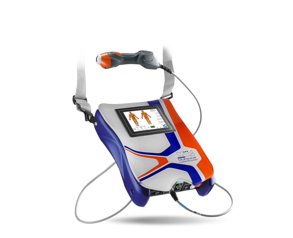
The following treatment protocols are designed for use with the ASA MLS Mphi 75 Laser, a dual-wavelength pulsed and continuous emission laser device, the HIKMICRO thermal imaging system for assessment, and a microcurrent (MENS) device with adjustable parameters. These protocols align with evidence-based practices while adapting to real-world clinical conditions.
4.1 Equipment Overview
Laser (ASA MLS Mphi 75):
- Wavelengths: 808 nm (continuous), 905 nm (pulsed)
- Peak power: up to 75W
- Preset programmes for chronic and acute conditions
- Emission modes: single-point, manual scan, trigger point
Microcurrent Unit:
- Adjustable current: < 600 μA
- Frequency range: 0.3–300 Hz
- Programmable for single or dual-channel use
- Biphasic waveform recommended for knee treatments
Thermal Imaging (HIKMICRO):
- Resolution: 240×240 SuperIR
- Refresh rate: 20 Hz
- Range: -20°C to 550°C
- Used for baseline assessment and monitoring inflammation zones
4.2 Protocol: Chronic Patellofemoral Osteoarthritis
Objective: Pain reduction, functional recovery, inflammation control.
Step 1 – Baseline Imaging
- Use thermal camera to identify localised hotspots (increased skin temperature suggests inflammatory areas).
- Document initial image for future comparison.
Step 2 – Laser Therapy (MLS Mphi 75)
- Mode: Chronic Pain Protocol
- Application Zones: Peripatellar region (6 points), medial and lateral joint line, suprapatellar pouch.
- Time: 1.5 min/point or 8–10 min in scanning mode.
- Energy Density: 4–6 J/cm² per point
- Sessions: 2–3 times per week for 3–4 weeks
Step 3 – Microcurrent Therapy
- Current: 100–200 µA
- Frequency: 0.5–10 Hz (chronic phase)
- Time: 20–30 min
- Placement: Electrode pads over medial/lateral capsule and vastus medialis
- Channels: Use 2 channels for bilateral stimulation
Step 4 – Kinesiotherapy
- Exercises: Seated leg extensions, mini squats, static bike, proprioception on unstable surface
- Reps/Sets: 3 sets of 10–15 reps depending on tolerance
- Progression: Add resistance after week 2 if tolerated
4.3 Protocol: Acute Flare-up or Post-Exercise Inflammation
Laser:
- Mode: Acute/Anti-inflammatory preset
- Time: 6–8 minutes (scanning over inflamed area)
- Energy Density: 2–3 J/cm²
- Frequency: Daily for 3 days, then taper
Microcurrent:
- Current: 40–80 µA
- Frequency: 0.3–1 Hz
- Time: 15–20 min
- Focus on vasodilation and ATP restoration
4.4 Considerations for Application
- Skin Contact: Ensure proper probe contact and alignment during laser.
- Pain Monitoring: Use VAS before/after to track progress.
- Thermal Feedback: Reassess thermal patterns weekly to monitor inflammatory reduction.
- Contraindications: Avoid active tumours, direct use over epiphyseal plates in adolescents, or in patients with pacemakers (for MENS).
4.5 Protocol Adjustments for Elderly Patients
Elderly patients often present with comorbidities, thinner skin, and altered healing capacity. Treatment should be adapted accordingly:
Laser:
- Mode: Chronic or Vascular protocol
- Energy: Begin with 3–4 J/cm²
- Frequency: 2×/week to reduce cumulative exposure
Microcurrent:
- Current: Lower initial dose (50–80 µA)
- Time: 15–20 min max
- Monitor hydration, medication interaction, and skin response
Kinesiotherapy:
- Type: Chair-based or aquatic options
- Emphasis: Flexibility, coordination, balance
- Load: Low to moderate intensity
5. Outcome Tracking and Monitoring
Continuous assessment ensures efficacy and guides protocol adjustments.
Thermal Imaging:
- Weekly thermographic scans to monitor changes in skin temperature.
- Reduction in hotspots correlates with inflammation resolution.
Pain Scale (VAS):
- Used before and after each session
- Helps gauge immediate analgesic effect
Range of Motion (ROM):
- Goniometric measurement of knee flexion/extension weekly
Functional Tests:
- Timed Up and Go (TUG), 6-Minute Walk Test (6MWT)
- Performed at baseline, mid-point, and discharge
Patient-Reported Outcomes:
- WOMAC or KOOS questionnaires
- Quality of life and joint-specific symptoms
6. Contraindications and Safety Measures
When applying combined therapies, it is essential to consider the specific contraindications and adhere to standard safety measures to ensure the well-being of patients.
6.1 Contraindications for Laser Therapy
- Active malignancy in the treatment area
- Pregnancy, especially over the abdominal and pelvic areas
- Direct application over the thyroid gland
- Epilepsy, particularly with pulsed modes
- Photosensitivity due to medication or systemic conditions
- Open wounds (unless indicated and guided by wound protocols)
- Over pacemakers or electronic implants
6.2 Contraindications for Microcurrent
- Pacemakers or defibrillators
- Pregnancy, unless medically approved
- Epileptic patients, especially with cranial placement
- Active bleeding or haemorrhage
- Over areas of thrombosis or suspected DVT
6.3 General Safety Guidelines
- Perform clinical assessment before initiating therapy
- Use protective eyewear for patient and therapist during laser application
- Monitor skin integrity before and after application
- Adjust settings according to age, skin tone, comorbidities
- Maintain hydration levels, particularly in elderly patients
- Document all parameters and outcomes for legal and clinical traceability
6.4 Practitioner Safety
- Follow manufacturer instructions for device calibration
- Ensure electrode and applicator hygiene
- Keep equipment certified and regularly maintained
6.5 Patient Education
- Inform patients about possible transient discomfort or delayed soreness post-treatment
- Advise to avoid extreme temperature exposure post-laser (e.g., cold showers)
- Encourage compliance with exercise and home care routines
Implementing these precautions not only improves therapeutic outcomes but also reinforces trust and confidence in the treatment process.
7. Conclusion and Future Directions
The integration of Low-Level Laser Therapy (LLLT), Microcurrent Electrical Neuromuscular Stimulation (MENS), and Kinesiotherapy offers a powerful, multimodal approach to treating knee osteoarthritis. This comprehensive methodology targets inflammation, pain modulation, tissue regeneration, and functional restoration — key pillars in the management of degenerative joint conditions.
Through this article, we have reviewed the scientific rationale, supported by credible studies, for combining these therapies. We’ve also presented detailed protocols tailored to different patient populations, including elderly individuals, and highlighted the importance of individualised adjustments to dosage, frequency, and progression. The use of diagnostic and tracking tools, such as thermal imaging and validated outcome measures, strengthens both clinical decision-making and the documentation of progress.
Importantly, the safety profile of this combined therapy model, when applied judiciously and within established contraindication guidelines, remains favourable — making it suitable for a wide demographic, from active adults to more vulnerable elderly patients.
Looking forward, future research should aim to explore larger-scale randomised controlled trials comparing the triple-modality approach with conventional mono-therapy interventions. There is also a promising opportunity to incorporate artificial intelligence and wearable technologies to enhance precision in treatment delivery and monitoring.
Ultimately, the combination of laser therapy, microcurrent, and movement rehabilitation is more than a therapeutic strategy — it represents a shift toward integrative, patient-centred care. As healthcare professionals seek to optimise outcomes in musculoskeletal health, this approach stands as a forward-thinking model grounded in evidence, technology, and human function.
ations of low-level laser therapy in physical therapy. Physical Therapy Reviews, 17(2), 94–99. https://doi.org/10.1179/1743288X11Y.0000000035
Gür, A., et al. (2003). Efficacy of low-level laser therapy in patients with knee osteoarthritis. Clinical Rheumatology, 22(2), 108–113. https://doi.org/10.1007/s10067-002-0677-1
Al Rashoud, A.S., et al. (2014). Efficacy of low-level laser therapy in the management of knee osteoarthritis: A systematic review and meta-analysis. Osteoarthritis and Cartilage, 22(11), 1757–1766. https://doi.org/10.1016/j.joca.2014.07.009
Cheng, N., et al. (1982). The effect of electric currents on ATP generation, protein synthesis, and membrane transport in rat skin. Clinical Orthopaedics and Related Research, (171), 264–272.
Curtis, D., Fallows, S.J., Morris, M., & McMakin, C. (2000). The efficacy of frequency specific microcurrent therapy on delayed onset muscle soreness. Journal of Bodywork and Movement Therapies, 4(1), 57–62. https://doi.org/10.1054/jbmt.1999.0169
Fransen, M., et al. (2015). Exercise for osteoarthritis of the knee: A Cochrane systematic review. British Journal of Sports Medicine, 49(24), 1554–1557. https://doi.org/10.1136/bjsports-2015-095424
Alayat, M.S.M., et al. (2017). Efficacy of laser therapy in the management of knee osteoarthritis: A systematic review and meta-analysis of randomised placebo or active-treatment controlled trials. Lasers in Medical Science, 32, 109–117. https://doi.org/10.1007/s10103-016-2083-2
Lopes-Martins, R.Á.B., et al. (2006). Effect of low-level laser therapy (LLLT) in the development of exercise-induced skeletal muscle fatigue and changes in biochemical markers related to postexercise recovery. Journal of Orthopaedic & Sports Physical Therapy, 36(12), 857–865. https://doi.org/10.2519/jospt.2006.2302
American College of Rheumatology. (2019). Guidelines for the Management of Osteoarthritis of the Knee and Hip. https://www.rheumatology.org/Practice-Quality/Clinical-Support/Clinical-Practice-Guidelines/Osteoarthritis
World Association for Laser Therapy (WALT) (2010). Recommended treatment doses for low-level laser therapy. https://waltza.co.za/documentation-links/recommendations/
Brosseau, L., et al. (2005). Therapeutic ultrasound for treating patellofemoral pain syndrome. Cochrane Database of Systematic Reviews, (1), CD003375. https://doi.org/10.1002/14651858.CD003375.pub2
Yousefi-Nooraie, R., et al. (2008). Low level laser therapy for osteoarthritis of the knee: A systematic review and meta-analysis. Canadian Medical Association Journal, 179(9), 997–1003. https://doi.org/10.1503/cmaj.080162
Delitto, A., et al. (2016). Physical therapist management of knee osteoarthritis: Clinical practice guidelines linked to the International Classification of Functioning, Disability, and Health. Journal of Orthopaedic & Sports Physical Therapy, 46(6), A1–A40. https://doi.org/10.2519/jospt.2016.0301
Ladeira, C.E. (2012). Evidence-based practice guidelines for management of osteoarthritis of the knee. Journal of Manual & Manipulative Therapy, 20(4), 208–212. https://doi.org/10.1179/2042618612Y.0000000017
